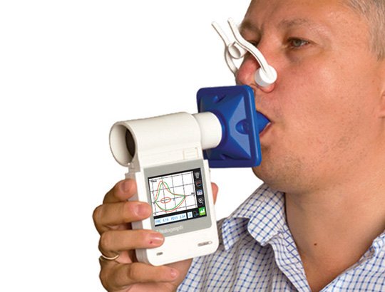
 Spirometer Market In the realm of respiratory health, Spirometer plays a vital role in assessing lung function and diagnosing various respiratory conditions. Whether having a known respiratory condition or simply want to monitor the lung health, understanding Spirometer is essential. In this comprehensive guide, we will delve into the world of Spirometer, exploring its purpose, procedure, and significance in maintaining optimal lung health. By gaining a deeper understanding of Spirometer, anyone can take a proactive approach to monitor the respiratory well-being and make informed decisions about the lung health. Spirometer is a diagnostic test that measures the volume and flow of air inhaled and exhaled by the lungs. It assesses lung function and helps healthcare professionals evaluate respiratory conditions such as asthma, chronic obstructive pulmonary disease (COPD), and other lung disorders. During a Spirometer test, the individual breathes into a handheld device called a spirometer, which measures the amount of air expelled and the rate at which it is exhaled. The results are then analysed to determine lung capacity, airflow limitations, and other parameters that aid in diagnosis and treatment planning. The Purpose and Benefits of Spirometer Spirometer serves multiple purposes in the field of respiratory health. It is used for diagnosing lung diseases, monitoring disease progression, assessing response to treatment, and evaluating lung function prior to surgery or medical procedures. By measuring lung volumes and airflow, Spirometer provides crucial information that helps healthcare professionals make accurate diagnoses and tailor treatment plans to individual patients. Regular Spirometer testing can provide valuable insights into lung health, enabling early detection of respiratory conditions and facilitating timely interventions. It helps individuals with chronic respiratory conditions monitor their lung function, detect changes, and make informed decisions about their treatment and lifestyle choices. Additionally, Spirometer can assist in assessing occupational lung diseases and evaluating the impact of environmental exposures on lung health. The Spirometer procedure involves a few simple steps. The individual takes a deep breath and exhales forcefully into the Spirometer mouthpiece. The process is repeated several times to ensure accurate and consistent results. The spirometer measures the volume of air exhaled and the speed of exhalation, generating a graphical representation of the airflow over time, known as a spirogram. To obtain accurate results, proper technique is crucial. The individual must fully exhale into the spirometer, blowing out as hard and as long as possible. Following specific instructions from a healthcare professional or Spirometer technician is essential to ensure accurate and reliable data. Spirometer results provide various measurements and values that assist in evaluating lung function. Key parameters include forced vital capacity (FVC), forced expiratory volume in one second (FEV1), FEV1/FVC ratio, and peak expiratory flow (PEF). These values are compared to predicted or normal values based on factors such as age, sex, height, and ethnicity. Abnormal Spirometer results may indicate airflow limitation, reduced lung capacity, or other respiratory abnormalities. They can help identify conditions such as asthma, COPD, restrictive lung diseases, or even early signs of lung damage. However, Spirometer results are just one component of a comprehensive diagnostic evaluation. Additional tests, clinical history, and physical examination are often necessary for a complete assessment and accurate diagnosis. Spirometer is a fundamental tool in assessing lung health and diagnosing respiratory conditions. By providing objective measurements of lung volumes and airflow, Spirometer assists healthcare professionals in evaluating lung function and tailoring treatment plans for individuals with respiratory diseases. Regular Spirometer testing can help detect early signs of lung damage, monitor disease progression, and guide therapeutic interventions. Understanding the purpose, procedure, and interpretation of Spirometer results empowers individuals to take an active role in their lung health and collaborate with healthcare professionals for optimal respiratory well-being. Read More - https://www.globenewswire.com/en/news-release/2021/08/16/2281170/0/en/Global-Spirometer-Market-to-Surpass-US-1-808-3-Million-by-2028-Says-Coherent-Market-Insights-CMI.html
0 Comments
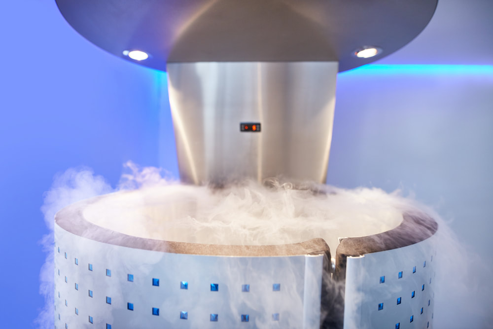 Cryotherapy Cryotherapy, the practice of exposing the body to extremely cold temperatures, has gained significant popularity in recent years. While it may seem like a trendy wellness fad, cryotherapy is rooted in science and offers various potential benefits. In this blog post, we will explore the science behind cryotherapy, how it works, and why it has become a trending treatment.
Cryotherapy involves subjecting the body to temperatures well below freezing, typically ranging from -110°C to -160°C (-166°F to -256°F), for a short duration, usually a few minutes. The most common method of Cryotherapy is whole-body cryotherapy (WBC), where individuals enter a specialized chamber that emits extremely cold air or liquid nitrogen. The process triggers several physiological responses in the body. Initially, the sudden cold exposure constricts blood vessels and reduces blood flow to the skin's surface. This protective mechanism is called vasoconstriction and aims to preserve core body temperature. As a result, the body redirects blood to vital organs and tissues, leading to increased oxygenation and nutrient delivery. Furthermore, the extreme cold activates the sympathetic nervous system, triggering the release of endorphins, adrenaline, and noradrenaline. These chemicals help reduce pain, improve mood, and increase energy levels. The potential benefits of cryotherapy have contributed to its rising popularity. While more research is needed, preliminary studies and anecdotal evidence suggest several positive effects. One of the most well-known benefits is pain relief. Cryotherapy may help reduce inflammation, alleviate muscle soreness, and provide relief for conditions like arthritis and fibromyalgia. Cryotherapy's impact on the immune system is another area of interest. Exposure to extreme cold temperatures may stimulate the production of anti-inflammatory cytokines and boost the body's natural defense mechanisms. This could potentially enhance immune function and help prevent illness. The release of endorphins during cryotherapy can also lead to improved mood and reduced symptoms of anxiety and depression. Additionally, the increase in metabolism and calorie expenditure during and after cryotherapy sessions may contribute to weight loss and improved metabolic function. Cryotherapy has gained traction among athletes and fitness enthusiasts due to its potential to enhance performance and aid in recovery. The cold exposure can help reduce post-exercise inflammation, accelerate muscle repair, and improve muscle function. The increasing popularity of Cryotherapy can be attributed to several factors. First, the desire for alternative and non-invasive wellness treatments has grown in recent years. Cryotherapy offers a unique approach that is quick, convenient, and appealing to those seeking holistic health solutions. Moreover, celebrities, professional athletes, and influencers have popularized cryotherapy by sharing their positive experiences and endorsing its benefits. This widespread exposure has generated curiosity and sparked interest in trying cryotherapy. The rising emphasis on self-care and wellness has also played a role in cryotherapy's popularity. People are more aware of the importance of taking care of their physical and mental well-being, and cryotherapy aligns with this trend by offering potential benefits for both. Cryotherapy's increasing popularity can be attributed to its unique scientific mechanisms and potential benefits. By triggering vasoconstriction, anti-inflammatory responses, and the release of endorphins, cryotherapy offers a range of advantages, including pain relief, muscle recovery, and metabolic boosts. The trendiness of Cryotherapy is also fueled by its endorsement from celebrities, athletes, and the convenience it provides in terms of time efficiency. However, it's important to note that cryotherapy may not be suitable for everyone, and individual experiences may vary. As research continues to delve into the science behind cryotherapy, it's clear that this innovative treatment is here to stay. Whether you're looking for a new approach to recovery, pain management, or overall well-being, cryotherapy. Read More - https://www.globenewswire.com/en/news-release/2021/12/07/2347543/0/en/At-9-CAGR-Global-Cryotherapy-Market-to-Surpass-US-452-0-Million-by-2028-Says-Coherent-Market-Insights-CMI.html 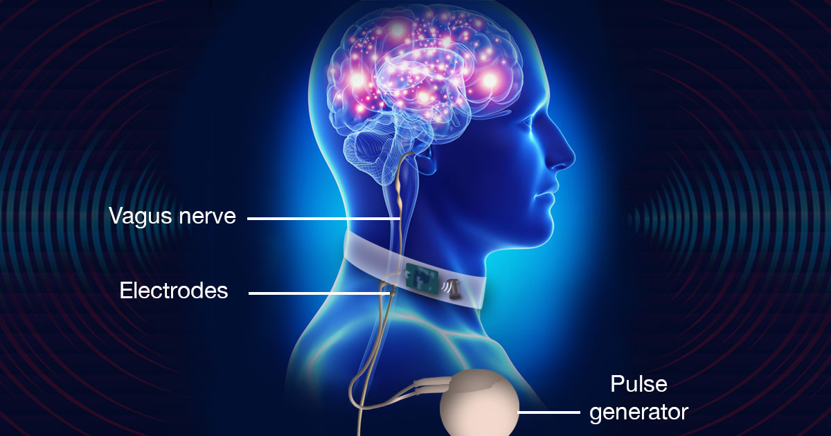 Vagus Nerve Stimulators Neurological disorders pose significant challenges to patients and healthcare professionals alike, often causing debilitating symptoms and impairing quality of life. In recent years, there has been growing interest and research into the potential of vagus nerve stimulators (VNS) as a therapeutic option for various neurological conditions. Vagus Nerve Stimulators is a technique that involves the electrical stimulation of the vagus nerve, a major nerve that extends from the brainstem to various organs throughout the body. By modulating the activity of the vagus nerve, VNS offers a promising avenue for treating neurological disorders and improving patient outcomes. One of the most well-known applications of VNS is in the management of epilepsy. Epilepsy is a chronic neurological disorder characterized by recurrent seizures. Traditional treatment approaches, such as medications, may not be effective for all patients and can have side effects. VNS provides an alternative treatment option, particularly for those who are resistant to medication. The electrical stimulation delivered by the Vagus Nerve Stimulators device helps regulate abnormal brain activity, reducing the frequency and severity of seizures. Numerous studies have demonstrated the efficacy of VNS in reducing seizure frequency and improving seizure control in epilepsy patients. Beyond epilepsy, VNS shows potential in treating other neurological disorders as well. Major depressive disorder (MDD) is a common mental health condition that can be challenging to manage. VNS has been investigated as a therapy for treatment-resistant depression, where traditional treatments have proven ineffective. By stimulating the vagus nerve, Vagus Nerve Stimulators is believed to modulate neurotransmitter systems involved in mood regulation, leading to improvements in depressive symptoms. Clinical trials have shown promising results, with some patients experiencing a significant reduction in depressive symptoms and an improved quality of life. In addition to epilepsy and depression, VNS is being explored as a treatment option for a range of other neurological conditions, including chronic pain, migraine, anxiety disorders, and even neurodegenerative diseases like Parkinson's and Alzheimer's. The potential benefits of VNS in these disorders stem from its ability to regulate neural circuits and modulate neurotransmitter release, offering a non-pharmacological approach with fewer systemic side effects. The utilization of Vagus Nerve Stimulators in treating neurological disorders is not without challenges. The selection of appropriate patients, determining optimal stimulation parameters, and understanding the long-term effects of VNS are areas of ongoing research. Furthermore, further studies are needed to elucidate the mechanisms of action and identify patient subgroups that are most likely to benefit from VNS therapy. However, the growing body of evidence supporting the efficacy and safety of VNS in neurological disorders provides hope for patients and healthcare professionals seeking alternative and effective treatment options. As research continues to advance, there is a need for collaboration between clinicians, researchers, and industry to refine Vagus Nerve Stimulators techniques, develop more advanced devices, and expand the range of neurological conditions for which VNS may be beneficial. By harnessing the potential of VNS, we may be able to transform the landscape of neurological disorder management and improve the lives of countless individuals worldwide. In conclusion, the exploration of vagus nerve stimulators in treating neurological disorders represents a significant advancement in the field of neuromodulation. Vagus Nerve Stimulators holds promise as an alternative or adjunctive therapy for various conditions, including epilepsy, depression, chronic pain, and more. While challenges and further research remain, the potential benefits of VNS in improving patient outcomes and enhancing quality of life cannot be overlooked. As our understanding of the complex interactions between the vagus nerve and neurological disorders deepens, VNS has the potential to revolutionize the treatment landscape and provide hope for individuals living with these challenging conditions.  Ultrasound Gels Ultrasound gels play a crucial role in medical imaging by enhancing the accuracy and clarity of ultrasound scans. These specialized gels provide a medium for efficient transmission of sound waves between the transducer and the patient's skin, facilitating optimal imaging results. The use of ultrasound gels has become a standard practice in various medical fields, including obstetrics, cardiology, and radiology, due to their significant benefits.
One of the primary functions of Ultrasound Gels is to eliminate air gaps between the transducer and the skin. When air is present, it creates an acoustic barrier that hinders the transmission of ultrasound waves and distorts the imaging quality. By applying a layer of gel, it fills in the gaps and ensures direct contact between the transducer and the skin. This direct contact allows for efficient sound wave transmission, resulting in clearer and more accurate images. Ultrasound Gels also act as a coupling agent, improving the transfer of ultrasound energy from the transducer to the patient's body. The gel's composition is designed to have similar acoustic properties to human tissue, minimizing the impedance mismatch between the transducer and the skin. This impedance matching enhances the efficiency of sound wave transmission and reception, leading to improved image resolution and detail. Moreover, ultrasound gels help to reduce the formation of artifacts in ultrasound images. Artifacts are undesired signals or distortions that can arise due to the presence of air or other substances that interfere with the ultrasound beam. The gel acts as a medium that ensures a consistent and uninterrupted flow of ultrasound waves, minimizing the occurrence of artifacts and improving the diagnostic accuracy of the images. Another significant advantage of Ultrasound Gels is their role in patient comfort and safety. The gel's lubricating properties facilitate smooth movement of the transducer over the patient's skin, reducing friction and discomfort. It also helps to maintain the appropriate temperature balance, preventing overheating or cooling of the skin during prolonged scanning procedures. Furthermore, ultrasound gels are generally water-based and hypoallergenic, minimizing the risk of skin irritation or allergic reactions, making them safe for use on a wide range of patients. Ultrasound gels are available in different viscosities to suit various imaging needs and preferences. Higher viscosity gels are often used for general imaging applications, while lower viscosity gels are suitable for more specialized procedures that require greater sensitivity, such as vascular or musculoskeletal ultrasound. The versatility of Ultrasound Gels allows healthcare professionals to customize their choice based on the specific imaging requirements of each patient and procedure. In addition to their role in traditional diagnostic imaging, ultrasound gels have found applications in therapeutic ultrasound procedures. In therapeutic ultrasound, focused sound waves are used to target specific areas for therapeutic purposes, such as pain management or tissue healing. The use of gels in these procedures helps to enhance the accuracy and effectiveness of the treatment by ensuring optimal sound wave penetration and delivery to the targeted area. In conclusion, ultrasound gels are essential tools in medical imaging, playing a vital role in enhancing the accuracy and clarity of ultrasound scans. By eliminating air gaps, improving sound wave transmission, reducing artifacts, and ensuring patient comfort and safety, these gels significantly contribute to the quality and diagnostic value of ultrasound images. Their versatility and wide range of applications make them indispensable in various medical fields. As technology continues to advance, the development of more advanced and specialized ultrasound gels is expected, further improving the efficiency and precision of ultrasound imaging. 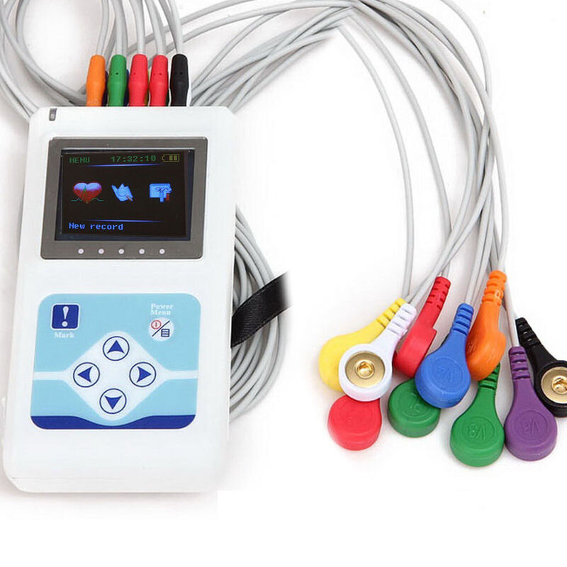 Holter Monitoring Systems Holter monitoring systems have emerged as a cornerstone in the field of cardiac health monitoring, revolutionizing the way we diagnose, track, and manage cardiovascular conditions. These systems enable continuous, non-invasive monitoring of a patient's heart rhythm and provide valuable insights into cardiac activity over an extended period.
By wearing a compact device equipped with electrodes, patients can go about their daily activities while the Holter monitoring system records and stores their heart's electrical signals. One of the key advantages of Holter Monitoring Systems is their ability to capture transient or intermittent cardiac events that may go undetected during short-term monitoring. Unlike traditional electrocardiography (ECG), which only captures a snapshot of heart activity, Holter monitoring systems offer a comprehensive and uninterrupted view of the heart's electrical patterns, allowing for more accurate detection and analysis of arrhythmias, palpitations, and other abnormalities. The future of cardiac health monitoring lies in the continued advancements of Holter Monitoring Systems. Technological innovations have led to the development of smaller, more comfortable devices that are easier for patients to wear for extended periods. Additionally, improved data storage capabilities and wireless connectivity enable seamless transfer of data to healthcare professionals, enhancing efficiency and reducing delays in diagnosis and treatment. Furthermore, Holter monitoring systems are becoming increasingly integrated with digital health platforms and remote patient monitoring systems. This integration allows for real-time monitoring and analysis of patient data, enabling healthcare providers to remotely assess a patient's heart rhythm, identify irregularities, and provide timely interventions. By leveraging these technological advancements, Holter monitoring systems are poised to play a pivotal role in telemedicine and remote healthcare delivery, particularly in underserved areas or for patients with limited access to healthcare facilities. The future of Holter Monitoring Systems also lies in their potential to be combined with other advanced technologies, such as artificial intelligence (AI) and machine learning algorithms. These intelligent algorithms can analyse the vast amounts of data collected by Holter monitors, detecting subtle patterns, predicting cardiac events, and providing personalized insights for each patient. This integration has the potential to enhance diagnostic accuracy, optimize treatment strategies, and enable more targeted and personalized care. Moreover, Holter Monitoring Systems are not limited to diagnosing arrhythmias alone. They can also provide valuable information about the effectiveness of medications, assess the impact of lifestyle changes, and aid in the management of chronic cardiovascular conditions. Continuous monitoring of a patient's heart rhythm over an extended period allows healthcare professionals to assess treatment response and make informed decisions to optimize therapy. In conclusion, Holter monitoring systems represent the future of cardiac health monitoring. With their ability to provide continuous, non-invasive monitoring and capture valuable data, these systems have transformed the way we diagnose and manage cardiovascular conditions. As technology continues to advance, Holter Monitoring Systems will become even more integrated, intelligent, and patient-centric, paving the way for enhanced personalized care, improved patient outcomes, and a healthier future for individuals with cardiac health concerns. 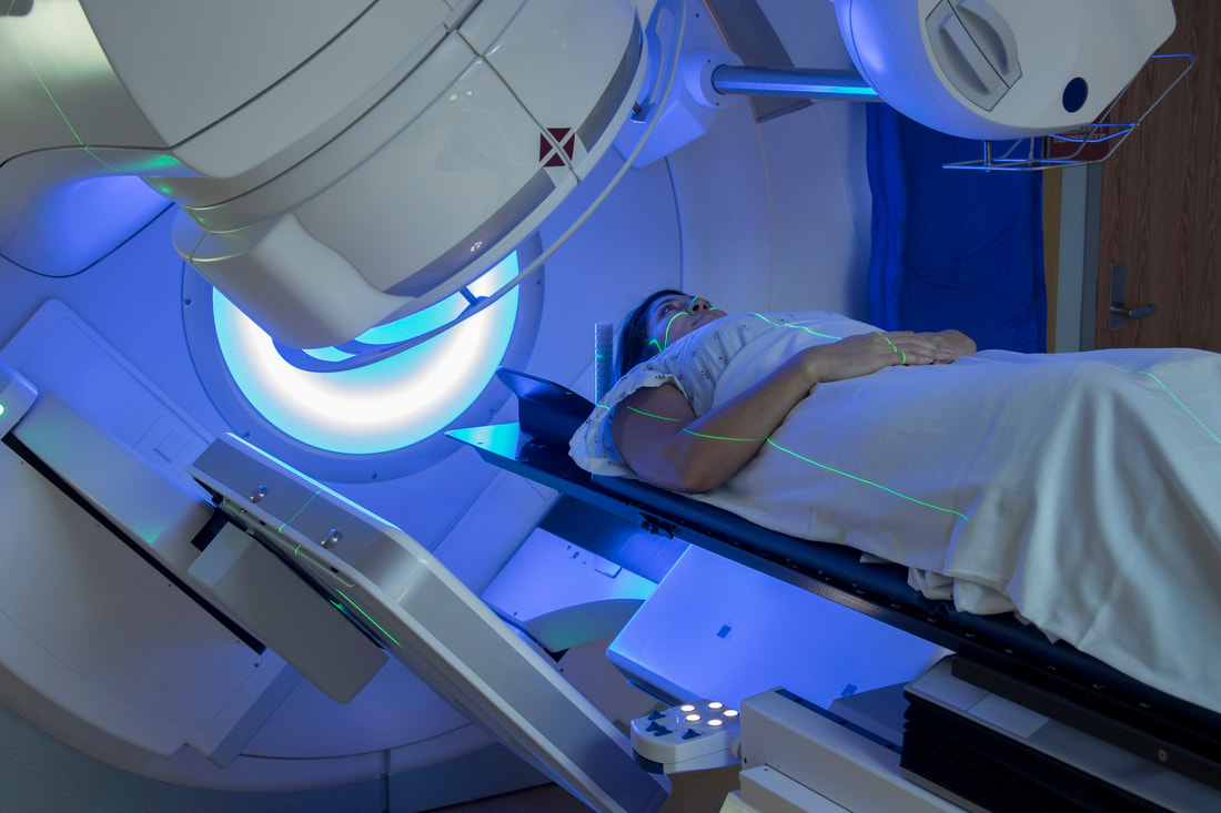 Radiotherapy Market Radiotherapy, often known as Radiotherapy or RT, is a treatment that uses ionising radiation to control or destroy cancerous cells. It is typically administered as part of cancer treatment and is frequently abbreviated as RT, RTx, or XRT. If a cancer is restricted to a single part of the body, Radiotherapy may be curative. It may also be used as adjuvant therapy to stop tumor recurrence following primary malignant tumor removal surgery (for example, early stages of breast cancer).
In tumors that are susceptible to chemotherapy, Radiotherapy has been utilised before, during, and after the chemotherapy. Radiation oncology refers to the branch of oncology that deals with Radiotherapy. Radiation oncologists are doctors who specialise in this field of medicine. As a result of its capacity to regulate cell proliferation, Radiotherapy is frequently administered to malignant tumors. Ionizing radiation kills cells by destroying the DNA of malignant tissue. Shaped radiation beams are directed from many angles of exposure to intersect at the tumor, producing a considerably higher absorbed dose there than in the surrounding healthy tissue, in order to spare normal tissues (such as skin or organs which radiation must pass through to treat the tumor). If the draining lymph nodes are clinically or radiologically associated with the tumor, or if there is a possibility of latent malignant dissemination, the radiation fields may additionally include those lymph nodes in addition to the tumor itself. The kind, location, and stage of the tumor, as well as the patient's general health, will determine the particular treatment intent (curative, adjuvant, neoadjuvant therapeutic, or palliative). Radiotherapy called total body irradiation (TBI) is used to get the body ready for a bone marrow transplant. Another type of Radiotherapy that reduces exposure to healthy tissue during procedures to treat malignancies of the breast, prostate, and other organs is brachytherapy, in which a radioactive source is put inside or close to the area that has to be treated. Several non-malignant illnesses can benefit from Radiotherapy, including trigeminal neuralgia, auditory neuromas, severe thyroid eye disease, pterygium, pigmented villonodular synovitis, and the prevention of keloid scar formation, vascular restenosis, and heterotopic ossification. Concerns regarding the possibility of radiation-induced tumors place restrictions on the use of Radiotherapy in non-malignant disorders. Radiotherapy has varying effects on the way various malignancies respond to it. The radio sensitivity of a cancer indicates how it will react to radiation. Modest radiation dosages quickly kill cancer cells that are highly radiosensitive. Leukemias, the majority of lymphomas, and germ cell cancers are among these. Most epithelial tumors are only moderately radiosensitive, necessitating much greater radiation doses to obtain a dramatic cure. Certain cancers are especially radio resistant, meaning that considerably greater doses than are likely safe in clinical practise are needed to provide a drastic cure. Radiotherapy is still a palliative option for many patients with metastatic melanoma despite the fact that radiation resistance is typically associated with renal cell carcinoma and melanoma. A current research topic that has shown some promise for treating melanoma and other cancers is the combination of Radiotherapy with immunotherapy. It is critical to distinguish between a tumor’s radio sensitivity, which is largely a laboratory measurement, and a cancer's radiation "curability" in actual clinical practise. Radiotherapy, for instance, typically cannot treat leukaemias since they have spread throughout the body. If lymphoma is limited to a single location of the body, it may be completely treatable. Similar to this, if a tumor is common and moderately radio responsive, it is frequently treated with curative dosages of Radiotherapy. For instance, non-melanoma skin cancer, cancers of the head and neck, breast cancer, non-small cell lung cancer, cervical cancer, anal cancer, and prostate cancer. Radiotherapy cannot cure metastatic tumors since it is impossible to treat the entire body, with the exception of oligometastatic disease. 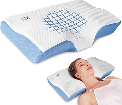 Cervical pillow Cervical Pillow are made to support the neck in the ideal position at the time of sleep and are precisely built to position the head and neck at the proper angles. The Cervical Pillow should rise where the neck will be and compress where the head will be at the time of sleep.
It's frequently referred to as a Cervical Pillow since the cervical spine refers to the upper portion of the backbone, which is where the neck is located. Comparatively speaking, a Cervical Pillow is much smaller than a regular bed pillow. This means that they are compatible with both vehicles and aircraft (or even boats!). It will support the head that is resting if wrap it around the neck. The first seven vertebrae of the spine make up the cervical spine. It supports the weight of the head, encloses and safeguards the spinal cord, and permits a variety of head motions. This is especially crucial when receiving physiotherapy since it supports treatment and speeds up healing. Cervical Pillow shouldn't be used for more than 20 minutes per day or as a pillow at night since they stretch or traction to the neck. Although it may take a person one to three days to become used to it, some people found it to be helpful and pleasant straight away. Pillows' primary function is to ease neck tension and improve head and neck posture. Extensor muscle endurance is increased in individuals with cervical spondylosis by ergonomic latex pillows. The pillow's height should be just right—not too high to cause the head to lean too far forward or too low to cause the neck to lengthen and the head to tilt upward. Although body type and preference are likely to affect pillow size, the cushion should typically remain between 4 and 6 inches tall to support the head and neck (and shoulders when lying on the back). Side sleepers need more support, as opposed to back and stomach sleepers. Memory foam could be used to make the Cervical pillow. The support provided by a memory foam pillow enables to sleep with the neck in its natural curve, which is the ideal posture for the spine. The first seven bones of the neck make up this region. Memory foam is mostly made of polyurethane, but it also contains other compounds that make it viscous and dense. It is frequently referred to as low-resilience polyurethane foam or "viscoelastic" polyurethane foam (LRPu). As the foam "cells" or bubbles are open, air may travel across them to form a matrix. Memory foam with a higher density softens in response to body heat, moulding to a heated body in just a few minutes. Newer foams could regain their original form faster. A cervical memory foam pillow's main function is to help persons who have problems maintaining a straight spine. Anybody can benefit from using a Cervical Pillow for neck pain relief and overall upright sleeping position. There isn't a better fit for side sleepers than cervical pillows. Neck pillows maintain optimal spinal alignment so that the muscles may unwind completely. Due to enhanced blood flow to the head, it also helps you sleep better by relaxing and supporting stiff muscles in the neck and shoulders. The design of cervical pillows, also known as orthopedic pillows, is unique. The pillow has a higher section for supporting the neck, and a lower region for supporting the head. The Cervical Pillow is the best in its class in terms of efficiency and toughness. It is a natural cushion that focuses on the natural healing of its users and is available with the washable cover. The design, size, and material options for Cervical pillows are varied. The pillow should support the neck as you sleep and be ergonomically built. Finding the ideal form for the particular body shape and size is crucial because no two bodies are the same. |
AuthorWrite something about yourself. No need to be fancy, just an overview. Archives
July 2023
Categories
All
|
 RSS Feed
